Ostatnia aktualizacja: 27/11/17
Wprowadzenie do uformowanych elementów krwi:
uformowane elementy to komórki, pozostałości komórek i fragmenty komórek we krwi. krwinki czerwone (RBC lub erytrocyty) stanowią ponad 95% uformowanych elementów.
![sekcja kapilary.](https://www.getbodysmart.com/wp-content/uploads/2017/10/Section-of-a-capillary-666x550.png)
![krwinka czerwona w części slajdu animacji naczyń włosowatych 2](https://www.getbodysmart.com/wp-content/uploads/2017/10/Red-blood-cells-in-the-blood-animation-slide-2-666x550.png)
![krwinka czerwona w części slajdu animacji naczyń włosowatych 3](https://www.getbodysmart.com/wp-content/uploads/2017/10/Red-blood-cells-in-the-blood-animation-slide-3-666x550.png)
![krwinka czerwona w części slajdu animacji naczyń włosowatych sekcja slajdu animacji kapilarnej 4](https://www.getbodysmart.com/wp-content/uploads/2017/10/Red-blood-cells-in-the-blood-animation-slide-4-666x550.png)
![krwinka czerwona w sekcji slajdu animacji kapilarnej 5](https://www.getbodysmart.com/wp-content/uploads/2017/10/Red-blood-cells-in-the-blood-animation-slide-5-666x550.png)
![krwinka czerwona w sekcji slajdu animacji kapilarnej 6](https://www.getbodysmart.com/wp-content/uploads/2017/10/Red-blood-cells-in-the-blood-animation-slide-6-666x550.png)
![krwinka czerwona w odcinku animacji naczyń włosowatych slajd 7](https://www.getbodysmart.com/wp-content/uploads/2017/10/Red-blood-cells-in-the-blood-animation-slide-7-666x550.png)
![krwinka czerwona w sekcji animacji naczyń włosowatych slajd 8](https://www.getbodysmart.com/wp-content/uploads/2017/10/Red-blood-cells-in-the-blood-animation-slide-8-666x550.png)
ponieważ brakuje im jądra i organelli, większość RBC we krwi nie jest w pełni funkcjonalnymi komórkami. Zamiast tego służą jako tymczasowe, wypełnione hemoglobiną pojemniki, które transportują tlen w organizmie.
również w utworzonych elementów jest pięć rodzajów białych krwinek (WBC lub leukocytów). Są częścią układu odpornościowego, który pomaga chronić organizm przed obcymi najeźdźcami. WBC są identyfikowane i klasyfikowane na podstawie ich poplamionego wyglądu.
trzy z WBC mają granulki cytoplazmatyczne i nazywane są granulocytami:
neutrofile
![odcinek kapilary.](https://www.getbodysmart.com/wp-content/uploads/2017/10/Section-of-a-capillary-666x550.png)
![neutrofil w części slajdu animacji kapilarnej 1](https://www.getbodysmart.com/wp-content/uploads/2017/10/Neutrophils-in-the-blood-animation-slide-1-666x550.png)
![neutrofil w części slajdu animacji kapilarnej 2](https://www.getbodysmart.com/wp-content/uploads/2017/10/Neutrophils-in-the-blood-animation-slide-2-666x550.png)
![neutrofil w części slajdu animacji kapilarnej 3](https://www.getbodysmart.com/wp-content/uploads/2017/10/Neutrophils-in-the-blood-animation-slide-3-666x550.png)
![a neutrofil w części slajdu animacji kapilarnej 4](https://www.getbodysmart.com/wp-content/uploads/2017/10/Neutrophils-in-the-blood-animation-slide-4-666x550.png)
![a neutrofil w części slajdu animacji kapilarnej 5](https://www.getbodysmart.com/wp-content/uploads/2017/10/Neutrophils-in-the-blood-animation-slide-5-666x550.png)
![a neutrofil w części slajdu animacji kapilarnej sekcja animacji kapilarnej slide 6](https://www.getbodysmart.com/wp-content/uploads/2017/10/Neutrophils-in-the-blood-animation-slide-6-666x550.png)
![neutrofil w sekcji slajdu animacji naczyń włosowatych 7](https://www.getbodysmart.com/wp-content/uploads/2017/10/Neutrophils-in-the-blood-animation-slide-7-666x550.png)
![neutrofil w sekcji slajdu animacji naczyń włosowatych 8](https://www.getbodysmart.com/wp-content/uploads/2017/10/Neutrophils-in-the-blood-animation-slide-8-666x550.png)
eozynofile
![sekcja kapilary.](https://www.getbodysmart.com/wp-content/uploads/2017/10/Section-of-a-capillary-666x550.png)
![An eosinophil in a section of capillary animation slide 1](https://www.getbodysmart.com/wp-content/uploads/2017/10/Eosinophils-in-the-blood-animation-slide-1-666x550.png)
![An eosinophil in a section of capillary animation slide 2](https://www.getbodysmart.com/wp-content/uploads/2017/10/Eosinophils-in-the-blood-animation-slide-2-666x550.png)
![An eosinophil in a section of capillary animation slide 3](https://www.getbodysmart.com/wp-content/uploads/2017/10/Eosinophils-in-the-blood-animation-slide-3-666x550.png)
![An eosinophil in a section of capillary animation slide 4](https://www.getbodysmart.com/wp-content/uploads/2017/10/Eosinophils-in-the-blood-animation-slide-4-666x550.png)
![An eosinophil in a section of capillary animation slide 5](https://www.getbodysmart.com/wp-content/uploads/2017/10/Eosinophils-in-the-blood-animation-slide-5-666x550.png)
![An eosinophil in a section of capillary animation slide 6](https://www.getbodysmart.com/wp-content/uploads/2017/10/Eosinophils-in-the-blood-animation-slide-6-666x550.png)
![Eozynofil w sekcji animacji kapilarnej slajdu 7](https://www.getbodysmart.com/wp-content/uploads/2017/10/Eosinophils-in-the-blood-animation-slide-7-666x550.png)
bazofile
![sekcja kapilary.](https://www.getbodysmart.com/wp-content/uploads/2017/10/Section-of-a-capillary-666x550.png)
![bazofil w sekcji slajdu animacji kapilarnej 1](https://www.getbodysmart.com/wp-content/uploads/2017/10/Basophils-in-the-blood-animation-slide-1-666x550.png)
![bazofil w sekcji slajdu animacji kapilarnej 2](https://www.getbodysmart.com/wp-content/uploads/2017/10/Basophils-in-the-blood-animation-slide-2-666x550.png)
![bazofil w sekcji slajdu animacji kapilarnej sekcja slajdów animacji kapilarnej 3](https://www.getbodysmart.com/wp-content/uploads/2017/10/Basophils-in-the-blood-animation-slide-3-666x550.png)
![bazofil w sekcji slajdów animacji kapilarnej 4](https://www.getbodysmart.com/wp-content/uploads/2017/10/Basophils-in-the-blood-animation-slide-4-666x550.png)
![bazofil w sekcji slajdów animacji kapilarnej 5](https://www.getbodysmart.com/wp-content/uploads/2017/10/Basophils-in-the-blood-animation-slide-5-666x550.png)
![bazofil w sekcji animacji kapilarnej slajdu 6](https://www.getbodysmart.com/wp-content/uploads/2017/10/Basophils-in-the-blood-animation-slide-6-666x550.png)
![bazofil w sekcji animacji kapilarnej slajd 7](https://www.getbodysmart.com/wp-content/uploads/2017/10/Basophils-in-the-blood-animation-slide-7-666x550.png)
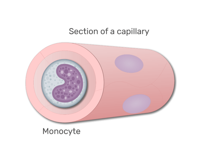
pozostałe dwa typy WBC nie mają granulek cytoplazmatycznych i są klasyfikowane jako agranulocyty:
limfocyty
![odcinek kapilary.](https://www.getbodysmart.com/wp-content/uploads/2017/10/Section-of-a-capillary-666x550.png)
![a limfocyty w sekcji slajdu animacji kapilarnej 1](https://www.getbodysmart.com/wp-content/uploads/2017/10/Lymphocytes-in-the-blood-animation-slide-1-666x550.png)
![a limfocyty w sekcji slajdu animacji kapilarnej 2](https://www.getbodysmart.com/wp-content/uploads/2017/10/Lymphocytes-in-the-blood-animation-slide-2-666x550.png)
![a limfocyty w sekcji slajdu animacji kapilarnej slajd animacji kapilarnej 3](https://www.getbodysmart.com/wp-content/uploads/2017/10/Lymphocytes-in-the-blood-animation-slide-3-666x550.png)
![a limfocyty w sekcji slajdu animacji kapilarnej 4](https://www.getbodysmart.com/wp-content/uploads/2017/10/Lymphocytes-in-the-blood-animation-slide-4-666x550.png)
![a limfocyty w sekcji slajdu animacji kapilarnej 5](https://www.getbodysmart.com/wp-content/uploads/2017/10/Lymphocytes-in-the-blood-animation-slide-5-666x550.png)
![a limfocyty w sekcji animacji kapilarnej slajdu 6](https://www.getbodysmart.com/wp-content/uploads/2017/10/Lymphocytes-in-the-blood-animation-slide-6-666x550.png)
![a limfocyty w sekcji animacji naczyń włosowatych slajd 7](https://www.getbodysmart.com/wp-content/uploads/2017/10/Lymphocytes-in-the-blood-animation-slide-7-666x550.png)
monocyty
Naucz się identyfikować komórki pod mikroskopem za pomocą tych quizów histologicznych i ćwiczeń znakowania.
![sekcja kapilary.](https://www.getbodysmart.com/wp-content/uploads/2017/10/Section-of-a-capillary-666x550.png)
![A monocyte in a section in capillary animation slide 1](https://www.getbodysmart.com/wp-content/uploads/2017/10/Monocytes-in-the-blood-animation-slide-1-666x550.png)
![A monocyte in a section in capillary animation slide 2](https://www.getbodysmart.com/wp-content/uploads/2017/10/Monocytes-in-the-blood-animation-slide-2-666x550.png)
![A monocyte in a section in capillary animation slide 3](https://www.getbodysmart.com/wp-content/uploads/2017/10/Monocytes-in-the-blood-animation-slide-3-666x550.png)
![A monocyte in a section in capillary animation slide 4](https://www.getbodysmart.com/wp-content/uploads/2017/10/Monocytes-in-the-blood-animation-slide-4-666x550.png)
![A monocyte in a section in capillary animation slide 5](https://www.getbodysmart.com/wp-content/uploads/2017/10/Monocytes-in-the-blood-animation-slide-5-666x550.png)
![A monocyte in a section in capillary animation slide 6](https://www.getbodysmart.com/wp-content/uploads/2017/10/Monocytes-in-the-blood-animation-slide-6-666x550.png)
![monocyt w sekcji w slajdzie animacji naczyń włosowatych 7](https://www.getbodysmart.com/wp-content/uploads/2017/10/Monocytes-in-the-blood-animation-slide-7-666x550.png)
![monocyt w sekcji w slajdzie animacji naczyń włosowatych 8](https://www.getbodysmart.com/wp-content/uploads/2017/10/Monocytes-in-the-blood-animation-slide-8-666x550.png)
małe utworzone elementy nazywane sąpłytki krwi (trombocyty). Są to fragmenty cytoplazmatyczne, które szczypają się z dużych komórek zwanych megakariocytami. Fosfolipidy uwalniane z płytek krwi pomagają zainicjować proces krzepnięcia.
![sekcja kapilary.](https://www.getbodysmart.com/wp-content/uploads/2017/10/Section-of-a-capillary-666x550.png)
![płytki krwi w części slajdu animacji naczyń włosowatych 1](https://www.getbodysmart.com/wp-content/uploads/2017/10/Platelets-in-the-blood-animation-slide-1-666x550.png)
![płytki krwi w części slajdu animacji naczyń włosowatych 2](https://www.getbodysmart.com/wp-content/uploads/2017/10/Platelets-in-the-blood-animation-slide-2-666x550.png)
![płytki krwi w części slajdu animacji naczyń włosowatych animacja slide 3](https://www.getbodysmart.com/wp-content/uploads/2017/10/Platelets-in-the-blood-animation-slide-3-666x550.png)
![płytki krwi w sekcji animacji naczyń włosowatych slide 4](https://www.getbodysmart.com/wp-content/uploads/2017/10/Platelets-in-the-blood-animation-slide-4-666x550.png)
![płytki krwi w sekcji animacji naczyń włosowatych slide 5](https://www.getbodysmart.com/wp-content/uploads/2017/10/Platelets-in-the-blood-animation-slide-5-666x550.png)
![płytki krwi w sekcji szkiełko animacji kapilarnej 6](https://www.getbodysmart.com/wp-content/uploads/2017/10/Platelets-in-the-blood-animation-slide-6-666x550.png)